Taking it seriously
I’ve always worked on the basis that medicine can’t always explain patients symptoms but that is usually because we don’t fully understand the nuances of the human body.I always recall one of mentors telling me about a heavy metal fan who came to outpatients complaining that everytime he started to ‘head bang’ he could feel something rattling inside his head! My colleague was a senior registrar in Neurology at the time and carried a full examination of this chap in his early 20’s. He found nothing of note on a thorough examination and concluded he shouldn’t shake his head vigorously and it wasn’t anything he was concerned about. This was in the mid 1980’s(!) when CT scans were in their infancy. A few years later this young man came in very ill with intractable headaches and confusion, as his third ventricle ependymoma had now grown to a size that was causing a physical obstruction to the flow of CSF ( the nourishing fluid that surrounds your brain/central nervous system) . The penny then dropped on what he was describing. This polyp like tumour was hanging in a natural fluid filled cavity in the middle of his brain and was swinging around ( like a punching ball) against the side of the cavity walls when he shook his head hard enough! It was a great lesson to be taught as a junior doctor; listen to your patients and take their symptoms seriously – even if you can’t make head or tail of it.
Thankfully these days imaging of the what is going on inside your head is much easier to get hold of. The current dangers are probably exposing patients to more radiation than necessary, so, clinical acumen is still important.

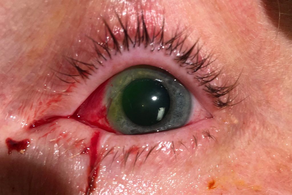
This week 2 patients came in under my care with clear clinical signs of optic nerve pathology. The first was a young lady who was pregnant and complained a gradual reduction of a gradual loss of vision in her eye over 5-6 months and intermittent headaches. Her vision was reduced in this eye, colour vision was reduced and examination of her pupils indicated a significant problem with light transmission down the right optic nerve. In these situations where I am particularly worried, I roll up to the neuro-imaging department and plea my case. As she was pregnant, we didn’t want to expose her to radiation from a CT scan and organised an MRI scan. Unfortunately the waiting list for an MRI scan is significantly longer than the wait for a CT scan, as the technology takes longer to obtain the images and the hospital has fewer machines because of the nature of the machines ( the MRI uses a massive magnetic to obtain the images and requires special housing) . I don’t think the radiology department was too impressed by her story although we were clearly worried about the hard signs we elicited. After a second visit to the department to chase this up, they took us a bit more seriously and moved her up the list a bit. The scan showed a satsuma sized benign tumour pressing on her right optic nerve. Interestingly this has grown because of the high levels of progesterone ( one of the sex hormones ) that occurs during pregnancy. So, off she has gone off to the neurosurgeons and obstetricians to decide if they need to shrink some of the tumour swelling with oral steroids, induce the delivery of the baby or remove her benign brain tumour. Currently , that is the order of the management strategy as decided by a multi-disciplinary meeting.
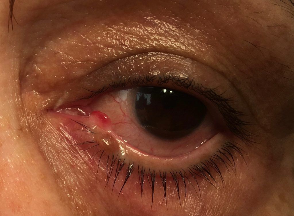
The second case is a man in his 20’s referred by his optician with reduced vision in one eye, reduced colour vision, a swollen optic disc, loss of his visual field in the affected eye and his pupils pointing towards reduced function in his optic nerve.These symptoms had gone on for 1-2 months before he decided to see his optician, who sent him straight up to the Emergency Eye Department.His scan showed something pressing on his optic nerve at the very back of his eye socket. The MRI couldn’t define what it is, but the most likely cause is an abnormal collection of blood vessels which had bled and is compromising the function of the nerve. He is getting more fancy CT images (CT angiography) to determine if there is blood flowing through this ‘shadow’. If there is , then probably this is best left alone, as any attempt to cut out blood vessels in this area is going to cause more collateral damage. It is just in a very awkward place.
The post Taking it seriously appeared first on Eric Barnes | Newcastle Eye Surgeon.